10 Questions You’ve Always Wanted to Ask About UFE
Alicia Armeli

If you have uterine fibroids and are nervous about treatment, you’re not alone. A recent survey revealed a staggering 90.5% of women with fibroids reported fears and concerns about their condition and their treatment options.1

To put some of these worries to rest, we sat down with Dr. Richard A. Reed, interventional radiologist at Huntington Hospital in Pasadena, Calif., to answer your top 10 questions about uterine fibroid embolization (UFE). It’s important to keep in mind that this blog doesn’t replace visiting your doctor.

 I heard UFE hurts. What type of pain can I expect during and after the procedure?

You’ll be under conscious sedation during the procedure. The pain occurs after the procedure and varies from woman to woman. The pain is typically crampy like a menstrual period and can be quite severe. It commonly begins minutes after the procedure is completed and is most intense the first night. This discomfort generally lasts one to three days. Medications are available to help control the pain.

What causes the pain after UFE?

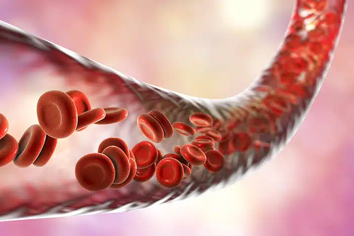
 UFE works by blocking blood flow to the fibroids, causing the tumors to die. This is accomplished by injecting an embolic agent, such as tiny microspheres, into the uterine arteries that supply fibroids with blood. Without a blood supply, fibroid tumors die, and pain occurs.

What is the embolic agent used for UFE and is it safe?

The embolic material I use in my practice is made up of tiny, biocompatible beads, meaning they don’t react with the body. This type of embolic material, Embosphere® Microspheres, has been used for over two decades in many types of embolization procedures in different parts of the body.

I’m worried these particles will travel to other places in my body, like my heart. Is that possible?

The embolic material is delivered to the uterine artery using a slim tube called a catheter. The tip of the catheter is positioned within the uterine artery and the particles travel via the blood flow to the uterus. The only way particles can go anywhere else in the pelvis is if the catheter tip isn’t in the correct position or if more embolic is injected than needed. If not done properly, reflux can occur, which means the particles move backwards and into other arteries in the pelvis, such as the bladder. This complication is very rare and has more to do with the experience of the doctor doing the procedure than the type of embolic.

I know I have a lot of fibroids. Can UFE treat each fibroid?

 UFE embolizes the entire uterus. By doing so, all of the fibroids should be treated. I’ve had many women ask why the uterus isn’t affected as well.  The reason UFE treats fibroids and doesn’t affect the uterus is because there’s collateral or additional blood flow from other vessels to the normal uterine tissue, allowing a doctor to selectively treat the fibroids while maintaining the viability of the uterus.

Is it safe for a fibroid to die inside of me, and what can I expect this to feel like?

Embolizing the fibroids causes them to shrink and die over time. This is what leads to symptoms improving. Before treatment, fibroids feel hard, which is why a common symptom is pelvic pressure. Once fibroids are treated, even without significant size change, they become softer. They no longer press on other organs or cause a woman’s belly to protrude. Fibroids can shrink to varying degrees—up to 50%.  Occasionally, fibroids will spontaneously die without any treatment. This may be seen on a magnetic resonance imaging (MRI) scan, and it’ll look the same as when images of fibroids are taken after UFE. There are typically no problems related to the presence of this type of fibroid in the uterus, whether it’s spontaneous or related to embolization. Fibroid expulsion, or the fibroid detaching from the uterus and passing from body, is an unusual occurrence post-UFE.

Is it necessary to have an MRI after UFE?

Many women will ask if an ultrasound will be sufficient, but an MRI provides crucial information—before and after the procedure.  An ultrasound shows the presence of masses, so you see where the fibroids are, but you can’t determine whether or not the treatment was a success. You also can’t tell if a woman has another condition with symptoms similar to fibroids called adenomyosis. With an MRI, doctors can get that information before the procedure to see if a woman is even a candidate for UFE. The 6-month follow-up MRI can show if all fibroids have been treated and the extent of the shrinkage. If a woman is having symptoms after UFE and the follow-up MRI shows viable fibroids, a repeat UFE may be helpful. On the other hand, if there are no viable fibroids on the follow-up MRI, I can counsel a woman that her symptoms most likely aren’t due to fibroids and that a repeat embolization won’t be helpful.

Will I stop getting my period after UFE?

A common reason women have UFE done is because of excessive bleeding during their menstrual cycle. The goal is to get their periods back to the way they were prior to having the severe bleeding. The majority of women will menstruate after the procedure, and it’ll come pretty close to the time they expect it. Any stress for women can cause the menstrual cycle to be a bit off, including the UFE procedure. It may take a few months after UFE for a woman’s cycle to regulate. There’s a small chance—under 10%—for women over 40 to go into early menopause after the procedure, causing them to stop menstruation all together.

Do I have to use birth control after UFE?

A woman having her cycle can become pregnant. If she wants to avoid pregnancy, then some kind of birth control should be used, for example condoms, an IUD, or birth control pills. Once a period comes back and is regular, it means a woman is ovulating, and she can become pregnant, so some kind of birth control is necessary if she wants to avoid that.

I want to have children. Is UFE the right treatment for me?

It’s possible for women to get pregnant following the procedure. But it’s controversial whether UFE is the right treatment for women who desire pregnancy. Traditionally, myomectomy—or the surgical removal of fibroids—has been the treatment of choice. There’s currently a larger body of research supporting myomectomy for women who have symptomatic fibroids and a strong desire to have children. There are randomized trials that suggest UFE is comparable to myomectomy for women who desire future fertility with more studies coming in the future.

Initially, UFE was only performed on women who weren’t interested in future fertility. These women were followed, and some became pregnant and did quite well with the pregnancies. Doctors realized that it is possible to conceive and deliver a healthy child. Over time, UFE was also offered to women who didn’t want to completely eliminate future fertility, as they would with hysterectomy, but wanted a less invasive procedure that has a high likelihood of eradicating fibroid-related symptoms.

It also depends on each individual woman. For someone young who wants to start a family, I would recommend myomectomy. And then there are women in their mid-to-late 40s who know they probably aren’t going to get pregnant but don’t want to completely eliminate future fertility with hysterectomy. These women really benefit from UFE.

The foregoing is not intended nor recommended as a substitute for medical advice, diagnosis or treatment. Always seek the advice of a qualified physician regarding any medical questions or conditions.

ABOUT THE DOCTOR Richard A. Reed, MD, is an interventional radiologist at Huntington Hospital in Pasadena, Calif. One of the first physicians in the US to perform UFE, Dr. Reed advocates for UFE public awareness and works to ensure women know all their fibroid treatment options.

 REFERENCES

  1. Knudsen, N. I., Wernecke, K. D., Siedentopf, F., et al. (2017). Fears and concerns of patients with uterine fibroids – a survey of 807 women. Geburtshilfe Frauenheikd, Sep;77(9):976-983.

Could Choosing Your Embolic Mean Less Post-UFE Pain?
Alicia Armeli

Minimally invasive uterine fibroid embolization (UFE) is a safe and effective treatment option for women with symptomatic uterine fibroids. In addition to avoiding surgery, UFE offers benefits that include low rates of serious complications, a faster recovery, an approximate 90% symptom improvement rate—and you get to keep your uterus.1

A drawback associated with UFE is cramping that patients reportedly experience within the first 24 hours after the procedure that often requires pain medication for relief.2 To help women be as comfortable as possible post-UFE, different pain management regimens have been designed.

But what if something as simple as choosing a specific type of embolic for UFE could make a substantial difference? Researchers at Saiseikai Shiga Hospital in Japan compared two embolics: Embosphere® Microspheres and gelatin sponge particles.3 They found that patients who underwent UFE with Embosphere Microspheres experienced significantly less post-procedural pain.

To understand why these results are so significant, we first need to understand the UFE procedure and what an embolic actually is. Under conscious sedation, a doctor called an interventional radiologist makes a nick in the wrist or groin area to gain access to the uterine arteries that supply fibroids with blood. A slim tube called a catheter is inserted and guided to these arteries. Once the catheter reaches the uterine arteries, tiny particles called embolic are released that then block blood flow to the fibroids. Deprived of blood, fibroids shrink over time and die, relieving women of fibroid-related symptoms, such as heavy menstrual bleeding and pelvic pain and pressure.

Although the UFE procedure is effective, the way it treats fibroids can also be a source of pain. To tackle this clinical problem from a different angle, Dr. Tetsuya Katsumori and a team of researchers decided to study how using different embolic material affected post-UFE pain in 101 patfients over a span of approximately eight years.3 Overall, 52 patients underwent UFE with Embosphere Microspheres and 49 patients were treated with gelatin sponge particles. Post-UFE pain was measured using a Visual Analog Scale where patients were able to choose a number that reflected the intensity of their pain. Dosages of pain and conscious sedation medications were also recorded.

Findings showed that not only were pain scores significantly lower among patients treated with Embosphere Microspheres within the first 24 hours, but hospitalization time was shorter and lower doses of pain and conscious sedation medications were required.3

Why did the patients treated with Embosphere Microspheres[ask_translation] experience less discomfort? The researchers noted that pain directly after UFE may be linked with blood restriction to normal uterine tissue. Uniform in size hydrophilic and spherical [ask_translation]Embosphere Microspheres better target the arteries that supply blood to the fibroids while still preventing severe blood restriction to normal uterine tissue.3 Gelatin sponge particles don’t have these same advantages. Given these outcomes, Katsumori and his team considered this information useful when selecting an embolic for UFE.

This advancement in UFE can aid doctors when choosing an embolic to use and—more importantly—can help women make informed decisions about their care. Because when it comes to fibroid treatment, the only place to be is in the know.

REFERENCES
1. Silberzweig, J. E., Powell, D. K., Matsumoto, A. H., et al. (2016). Management of uterine fibroids: a focus on uterine-sparing interventional techniques. Radiology, Sep;280(3):675-692.

2. Kim, H. S., Czuczman, G. J., Nicholson, W. K., et al. (2008). Pain levels within 24 hours after UFE: A comparison of morphine and fentanyl patient-controlled analgesia. Cardiovasc Intervent Radiol, Nov-Dec;31(6):1100-1107.

3. Katsumori, T., Arima, H., Asai S., et al. (2017). Comparison of pain within 24 h after uterine artery embolization with tris-acryl gelatin microspheres versus gelatin sponge particles for leiomyoma. Cardiovasc Intervent Radiol, Nov;40(11):1687-1693.

Radiologists Travel to Kenya to Provide Women with Life-Saving UFE


It’s well documented in medical literature that women of African descent develop uterine fibroids five to six years earlier and are more than 5 times as likely to have severe symptoms than Caucasian women of a similar family history.1

With the increasing number of women requiring fibroid treatment, hospitals in Kenya have welcomed international doctors to treat their patients with uterine fibroid embolization (UFE).

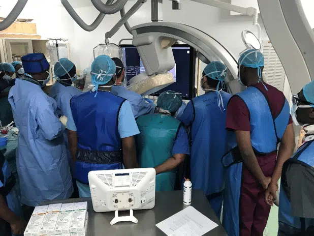
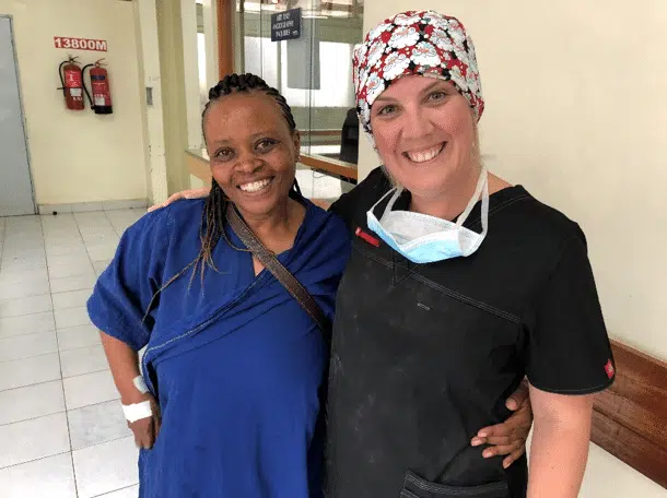
In July 2018, Dr. Darren Klass, interventional radiologist with Vancouver Coastal Health in Vancouver, British Columbia, Canada, traveled to Kenya with Merit Medical Systems Inc.—sponsor of Ask4UFE—to educate Kenyan gynecologists and radiologists on this uterine-sparing, minimally invasive option. By providing one-on-one UFE training to gynecologists—the doctors responsible for referring patients to radiologists for treatment—they can start recommending and providing this option to their patients.

In an intensive 10 days, Dr. Klass hosted a number of UFE training courses to several clinical teams in Karen and Kenyatta National Hospital in Nairobi and Aga Khan in Mombasa. Patients were also scheduled to undergo UFE in both cities. To help make this treatment possible, Merit Medical donated a portion of the embolic particles used for UFE procedures, called Embosphere® Microspheres. Dr. Klass observed and trained the clinical teams while they treated their own patients.

Traditionally, UFE has been performed through the femoral artery in the groin area. However, UFE can now be done via the radial artery in the wrist—a technique known as transradial access. This approach proved to be an eye-opener to both the Kenyan interventional radiologists and their clinical teams. Doctors observed how their patients were able to move and walk immediately post UFE and how patients could make themselves comfortable without having to lay still on their backs, which is required with femoral UFE.

All of the clinical skills taught and the education provided proved especially useful on the eve of the final day in Mombasa. Following a long day of training, Dr. Klass was called by a gynecologist to discuss an emergency case. A bedside consultation was done together with the referring gynecologist at around 8pm. The patient underwent a successful UFE procedure the following day and was discharged 24 hours post UFE. In this particular case, due to extenuating patient circumstances, UFE provided not only a uterine-sparing therapy for symptomatic fibroids but also a life-saving option. This experience was more than enough to show referring gynecologists that UFE is an effective fibroid treatment option. The gynecologist presented the case to an audience of doctors at a weekly Kenyan Medical Association meeting the night following the procedure.

The skills learned by the Kenyan radiologists can have a far-reaching impact, even beyond UFE. Because of the training with Dr. Klass, radiologists in both Mombasa and Nairobi can now also perform embolization for post-partum hemorrhage. Statistics show that 6,300 women die in Kenya each year during pregnancy and childbirth.2 A 2015 Kenyan policy document reported the maternal mortality rate at over 2,000 per 100,000 live births in North Eastern Kenya, and 212 per 100,000 live births in Nairobi, with hemorrhage accounting for 44% of those deaths.2

“Post-partum hemorrhage is an avoidable complication of childbirth and providing the radiologists here with the ability to treat this devastating complication with an effective therapy will hopefully have a dramatic impact on this shocking statistic,” Dr. Klass explains.

The time spent in Kenya was brief, but the main objective was achieved. Kenyan doctors can now provide these essential services to women.

“I arrived here wanting to improve the lives and care of women in Kenya,” Dr. Klass tells Ask4UFE. “I am pleased that by training radiologists in Kenya to perform this life-changing procedure, I saw this happening. Every single person I met in Kenya was grateful for the purpose of this trip.”

REFERENCES
1. Huyck, K. L., Panhuysen, C. I., Cuenco, K. T., et al. (2008). The impact of race as a risk factor for symptom severity and age at diagnosis of uterine leiomyoma among affected sisters. Am J Obstet Gynecol, Feb;198(2):168.e1-9.
2. National Council for Population and Development. (2015, Jun). Reducing Maternal Deaths in Kenya (Policy Brief No. 46). Retrieved from http://www.ncpd.go.ke/wp-content/uploads/2016/11/Policy-Brief-46-Maternal-Deaths-in-Kenya-1.pdf.

UFE and Myomectomy Work Together to Treat Rare Fibroids
Alicia Armeli

Intracavitary uterine fibroids. Try saying that five times fast. Besides being difficult to pronounce, this type of fibroid that grows within the uterine cavity may also be a challenge to treat. Like other fibroids, intracavitary fibroids aren’t cancerous, but they can be the cause of horrendous symptoms like prolonged, heavy periods, anemia, and extreme fatigue. Ugh.

Each year, hundreds of thousands of women seek treatment to find relief from the pain and suffering they experience with uterine fibroids.1 But if you’re a woman with intracavitary fibroids, you may not be a candidate for some of the most effective minimally invasive treatment options.2 So what’s a girl to do?

Before the word hysterectomy (a surgery that removes the entire uterus) crosses your mind, new research says there may be another way. A study conducted at University of California Irvine Medical Center in Orange, Calif., found that using uterine fibroid embolization (ufe) and hysteroscopic myomectomy as a combination therapy may be a safe and effective uterine-sparing option for women who have intracavity fibroids.2

Treating intracavitary fibroids with only ufe has been linked with a higher complication rate and may be a problem for women with large fibroids.2,3 Successfully treating fibroids with myomectomy alone depends much on where fibroids are located as well as their size, which may make intracavitary fibroids a challenge to treat.2

ufe is a nonsurgical procedure performed by an interventional radiologist and works by blocking the vessels that supply blood to the fibroids, causing them to shrink and symptoms to improve. Hysteroscopic myomectomy is performed by a gynecologist and involves surgically removing fibroids through the vagina and cervix.

To investigate how these two therapies work together, researchers examined treatment among 10 women with intracavitary fibroids who wanted to avoid hysterectomy.2  Each patient underwent the ufe procedure and then hysteroscopic myomectomy was planned for approximately one to two months later.

Results showed a 90% clinical success rate.2  One patient expulsed, or passed a fibroid, six days after ufe which was then removed, and one patient decided against myomectomy because she remained symptom-free after ufe. Around two months later, five patients underwent successful hysteroscopic myomectomy to remove any avascular fibroids left over after ufe. Two patients were scheduled for surgery at a later date. The remaining patient underwent a hysterectomy because her symptoms didn’t improve after the combination treatment. No short-term complications were seen.

For many women, finding a treatment option that avoids taking drastic measures like a hysterectomy is just as important as getting rid of bothersome symptoms. And although more research is needed, this study can provide hope for women suffering from intracavitary fibroids, offering a potential way to avoid hysterectomy while still finding relief.

REFERENCES

  1. US Department of Health and Human Services, National Institutes of Health. (2016). How many people are affected by or at risk of uterine fibroids? Retrieved from https://www.nichd.nih.gov/health/topics/uterine/conditioninfo/people-affected
  2. Khalsa,B. Costantino , M., &Goodwin, S. (2017). Uterine artery embolization followed by elective myomectomy for the treatment of intracavitary fibroids: Preliminary experience. J Vasc Interv Radiol, Feb;28(2):S47.
  3. Spies, J. B. (2016). Current role of uterine artery embolization in the management of uterine fibroids. Clin Obstet Gynecol, Mar;59(1):93-102.
This One Tool Could Help Predict Fibroid Growth
Alicia Armeli

Wouldn’t it be great if there were a tool that could help predict how much and how fast your fibroids grow? Keep reading.

A study by researchers at the VU University Medical Center in Amsterdam, The Netherlands, found that by measuring the vascular index of a fibroid, also known as its vascularization or the amount of blood vessels within the fibroid tissue, they could predict how much and how fast a fibroid would grow a year into the future.1

Between March 2012 and March 2014, 66 premenopausal women diagnosed with a max of two fibroids 8 cm or smaller participated in 1 year of follow-up care without undergoing fibroid treatment.1 The researchers used a three-dimensional (3D) ultrasound with power Doppler, a tool that can detect blood flow in vessels, especially those found inside organs. To measure the ongoing fibroid vascular index, each woman had a 3D ultrasound with power Doppler at the start of the study and then at 3, 6, and 12 months. Change in fibroid volume and fibroid growth rate were also calculated.

Results showed that fibroid volume and growth rate were related to its initial vascular index.1 Over 12 months, the average fibroid growth rate was 8.98%. But for fibroids that were highly vascularized, or in other words had more blood vessels present, the fibroid growth rate was greater and detected at 10.5%. If a fibroid’s vascular index increased by just 1%, it was linked with a 7-cm3 larger fibroid volume at the 1-year mark.

How can determining fibroid growth better the care women receive?

Uterine fibroids are noncancerous tumors that grow in the wall of the uterus. They can be the cause of heavy periods, pelvic pain and pressure, painful sex, and infertility. Predicting fibroid growth, especially among women with fast-growing fibroids, can help doctors narrow down which treatments could be the most effective for each individual.

For example, if fast-growing fibroids are detected early, medications that prevent fibroid growth might be helpful to these women.2 Among those who desire pregnancy, fast-growing fibroids that could potentially impair fertility can be detected and treated early on.2 Even if a woman has fibroids and isn’t having symptoms, predicting their growth rate can help in creating an effective follow-up care plan.2

Using fibroid vascularization as a tool can especially be helpful when treating women with minimally invasive procedures like uterine fibroid embolization (UFE). UFE works by targeting and blocking blood flow to the fibroids, causing them to shrink and symptoms to improve. By investigating which fibroids are highly vascularized and which aren’t may be helpful in determining how effective UFE will be, as UFE success may be limited among fibroids that aren’t vascularized.2

According to the researchers, we don’t yet have a way to strongly predict fibroid growth that can be used in everyday practice.1 Although this method of fibroid care seems promising, more research is needed to confirm the results of this study. Because of this, coverage by health insurance is not yet determined. But with additional data, predicting fibroid growth could become more economical and accessible to women, helping them to receive the best treatment and avoid unnecessary therapies.

REFERENCES

  1. Nieuwenhuis, L. L., Keizer, A. L., Stoelinga, B., et al. (2018). Fibroid vascularisation assessed with three-dimensional power Doppler ultrasound is a predictor for uterine fibroid growth: A prospective cohort study. BJOG, Apr;125(5):577-584.
  2. Dueholm, M. (2018). Fibroid vascularisation as a predictor for uterine fibroid growth. BJOG, Apr;125(5):585.
Anxious? Why Women Worry More Than Men & What We Can Do About It.
Alicia Armeli


A bit of anxiety every now and then is part of being human. For many of us, worrying and uneasiness are only temporary. But if you suffer from an anxiety disorder, you’re probably all too familiar with that nagging feeling that lasts for months, fears that get bigger as time goes on, and how all of this interferes with your everyday life.

As women, research shows, we’re almost twice as likely as men to get an anxiety disorder in our lifetime.1 The million-dollar question is why.

“In its most simple form, anxiety is the experience of tension between you and the present moment,” explains Katherine Schafler, LMHC, NYC-based women’s emotional health expert. And it can come in many forms, including generalized anxiety disorder (GAD), panic disorder, social anxiety disorder, obsessive compulsive disorder (OCD), and post-traumatic stress disorder (PTSD). Although anxiety doesn’t discriminate from one person to the next, it does seem to affect women more.

Fluctuations in sex hormones have been tagged as a possible cause for our boost in anxiety. But according to Margaret Altemus, MD, Director of the Payne Whitney Women’s Program at Weill Medical College Department of Psychiatry, we don’t know exactly why anxiety differs between women and men.2

“It doesn’t seem to be related to estrogen,” Dr. Altemus said in an Anxiety and Depression Association of America podcast. “In children, girls are twice as anxious as boys, so it’s even before the menstrual cycle starts that you see sex differences.”2 Being susceptible to anxiety, she explained, could go as far back as hormone levels in utero that may affect a developing fetus. Other possibilities that could contribute to the gender anxiety gap are major reproductive events and illnesses. Childbirth and the postpartum period have been seen to increase anxiety in women, as have reproductive diseases, such as uterine fibroids and endometriosis.3,4,5

And as if being from Mars and Venus weren’t enough, a woman’s brain chemistry differs greatly from that of a man’s, as do our coping mechanisms. When women face stress, they’re more likely to ruminate about their problems, writes Olivia Remes, anxiety researcher and doctoral candidate at the University of Cambridge in England.6 This can amplify anxiety. In contrast, she explains, men engage more in active, problem-focused coping.

Data has also shown that girls and women are more likely to encounter physical or mental abuse, and this experience is a known risk factor for PTSD. Childhood abuse may also change brain chemistry, affecting individuals as adults, predisposing them to anxiety disorders.7

Given all the potential associations and what-ifs, it’s clear more research needs to be done on behalf of our anxious minds. Science may not yet understand the connection between women and anxiety, but the good news is it’s treatable.

“When anxiety is ongoing, you may experience regular trouble sleeping, eating, and/or socializing. These are immediate signs that you need to get professional help,” Schafler tells Ask4UFE. It depends highly on the individual, she notes, but therapeutic interventions can include talk therapy, psychotropic medication, support groups, assertiveness training, animal-assisted therapy, and mindfulness exercises.

Women can also play an active role in their own treatment and engage in positive lifestyle changes like incorporating regular exercise, a healthy diet, and keeping positive relationships—this includes the relationship we have with ourselves.

“Surround yourself with people, places, and things that help you feel a sense of expansion, as opposed to contraction,” Schafler suggests. “Just as important as understanding who you are is understanding what you need.”

And never hesitate for one second to ask for it.


ABOUT THE THERAPIST  Katherine Schafler, LMHC, is an NYC-based women’s emotional health expert. With dual master’s degrees in clinical assessment and psychological counseling from Columbia University and post-graduate training and certification from the Association for Spirituality and Psychotherapy in NYC, Katherine helps women change their lives for the better through counseling and tailoring her approach to meet the unique needs of each of her clients. You can find her at https://www.katherineschafler.com  

REFERENCES

  1. Anxiety and Depression Association of America. (n.d.). Women and Anxiety. Retrieved from https://adaa.org/find-help-for/women/anxiety#
  2. Anxiety and Depression Association of America. (Producer). (n.d.). Anxiety Disorders in Women [Audio podcast]. Retrieved from https://adaa.org/learn-from-us/from-the-experts/podcasts/anxiety-disorders-women
  3. Shlomi, P. I., Huller, H. L., Baum, M., et al. (2014). Postpartum anxiety in a cohort of women from the general population: Risk factors and association with depression during last week of pregnancy, postpartum depression, and postpartum PTSD. Sr J Psychiatry Relat Sci, 51(2):128-134.
  4. Ghant, M. S., Sengoba, K. S., Recht, H., et al. (2015). Beyond the physical: A qualitative assessment of the burden of symptomatic uterine fibroids on women’s emotional and psychosocial health. J Psychosom Res, May;78(5):499-503.
  5. Friedl, F., Riedl, D., Fessler, S., et al. (2015). Impact of endometriosis on quality of life, anxiety, and depression: An Austrian perspective. Arch Gynecol Obstet, Dec;292(6):1393-1399.
  6. Remes, O. (2016, Jun 10). Women are far more anxious than men—here’s the science. Retrieved from https://theconversation.com/women-are-far-more-anxious-than-men-heres-the-science-60458
  7. Harvard Medical School. (2002). Coping with Anxiety and Phobias. Retrieved from http://hrccatalog.hrrh.on.ca/InmagicGenie/DocumentFolder/copinganxietyphobias.pdf
These 3 STDs Are on the Rise (…and Are Becoming More and More Difficult to Treat)

More than 1 million sexually transmitted diseases (STDs) are acquired every day worldwide.1 If you’re sexually active, STDs are a common fact of life. So common, in fact, that one in two sexually active Americans will get an STD by age 25.2

But an alarming trend is emerging. Three specific STDs are on the rise. According to the latest annual Centers for Disease Control and Prevention’s (CDC) Sexually Transmitted Disease Surveillance Report, more than two million cases of chlamydia, gonorrhea, and syphilis were reported in the US in 2016—a record high.3

The majority of the new diagnoses—a staggering 1.6 million—were cases of chlamydia, with young women making up nearly half of all infections reported.3 Closing in at second place was gonorrhea with 470,000 cases.3  Behind chlamydia, gonorrhea is the second most commonly reported disease in the US.4 And last, primary and secondary syphilis—the most infectious stages of the disease—had a reported 28,000 cases.3  Since 2015, chlamydia, gonorrhea, and syphilis have increased by 4.7%, 18.5%, and 17.6%, respectively.4

Superbugs & Antibiotic Resistance

Chlamydia, gonorrhea, and syphilis are infections caused by bacteria. So why not knock them out with a good ol’ fashioned dose of antibiotics? Generally, these STDs can be treated with antibiotics, but when they aren’t taken correctly (ahem…you didn’t follow your doctor’s instructions) or when they’re used too much, the result is a superbug that has evolved to protect itself from the effects of medications, making it resistant to treatment.5

Gonorrhea has developed the strongest antibiotic resistance with some strains of bacteria not even responding to any available antibiotics, whereas antibiotic resistance in chlamydia and syphilis isn’t as common but does exist.5 According to the CDC, this complicates successfully treating diseases like gonorrhea because there aren’t many effective drugs available that are both highly researched and tolerated.6

“Increases in STDs are a clear warning of a growing threat,” said Jonathan Mermin, MD, MPH, director of the CDC’s National Center for HIV/AIDS, Viral Hepatitis, STD, and TB Prevention in a 2017 press release. “STDs are a persistent enemy, growing in number, and outpacing our ability to respond.”3

How Does This Affect Me?

Both chlamydia and gonorrhea affect women disproportionately because early infection may not show symptoms.4

Left untreated, each disease comes with serious health complications.4  Both chlamydia and gonorrhea can move into the upper reproductive tract—such as the uterus and fallopian tubes—and lead to pelvic inflammatory disease (PID). PID increases the risk of infertility, ectopic pregnancy, pelvic pain, and is linked to chronic conditions like uterine fibroids. Women can also pass chlamydia onto their newborns during delivery, which can lead to blindness and pneumonia. A woman with untreated syphilis is at a greater risk for stillbirth and infant death. What’s also alarming is if you’re infected with chlamydia, gonorrhea, or syphilis, you’re two to three times more at risk of contracting HIV.5

What’s Being Done?

Responding to the threat of antibiotic resistance, the World Health Organization (WHO) released new recommendations for health professionals to follow that reflect the most effective antibiotic treatments of these STDs.5

Both WHO and the CDC encourage national, state, and local health departments to track the patterns of antibiotic resistance to improve detection of the diseases. Safe sex practices, like using condoms and having open dialogue about STDs within the community, are also recommended.3,5

What If I Get Infected with Chlamydia, Gonorrhea, or Syphilis?

Don’t freak out just yet. Breathe. As mentioned, chlamydia and syphilis most likely can still be treated with antibiotics. If you’re infected with gonorrhea, the current treatment recommendation is what’s referred to as “dual therapy” or using two types of drugs together. This involves a single shot of ceftriaxone and azithromycin taken by mouth. According to the CDC, taking the medication as prescribed will stop the infection.7

What Can I Do to Protect Myself?

The wise, old saying goes something like this: An ounce of prevention is worth a pound of cure. That being said, STDs are nothing to be ashamed of, but it only takes one unprotected sexual encounter to acquire an STD. There are many things you can do to prevent STDs and keep you and your partner(s) healthy.8

  • Use a latex condom correctly. This means every time you have anal, vaginal, and oral sex.
  • Talk about it. STDs are nothing to be embarrassed about. Talk openly with your partner(s) before having sex. Discuss any questions or concerns you may have with your doctor.
  • Have fewer sexual partners. Reducing your number of sex partners can lower your risk of STDs.
  • Mutual monogamy. Engaging in a long-term mutually monogamous relationship with an uninfected partner is a dependable way to avoid STDs.
  • Get tested regularly. And require that your partner(s) get tested, too. Remember, many STDs may not have symptoms but can still result in serious health consequences. Share your test results with your partner(s).
  • Get vaccinated. For STDs like the human papillomavirus (HPV) and hepatitis B, vaccines are a safe and effective way to help you avoid health problems associated with the infections.
  • Abstinence. Those dry spells aren’t necessarily a bad thing. The most reliable way to prevent infection is to not have sex.

This month is National STD Awareness Month. Pledge today to practice safe sex by joining the CDC’s Treat Me Right Campaign. Become aware of the ways you can keep yourself safe and talk to your doctor about the care you need to be sexually active and healthy:

https://www.cdc.gov/std/sam/index.htm?s_CID=tw_STD0180189


REFERENCES

  1. World Health Organization. (2016 Aug). Sexually transmitted infections (STIs). Retrieved from http://www.who.int/mediacentre/factsheets/fs110/en/
  2. American Sexual Health Association. (n.d.). Statistics STIs. Retrieved from http://www.ashasexualhealth.org/stdsstis/statistics/
  3. Centers for Disease Control and Prevention. (2017, Sep 26). STDs at record high, indicating urgent need for prevention. Retrieved from https://www.cdc.gov/media/releases/2017/p0926-std-prevention.html
  4. Centers for Disease Control and Prevention. (2017, Sep 26). 2016 Sexually Transmitted Disease Surveillance Report. Retrieved from https://www.cdc.gov/std/stats16/toc.htm
  5. World Health Organization. (2016, Aug 30). Growing antibiotic resistance forces updates to recommended treatment for sexually transmitted infections. Retrieved from http://www.who.int/mediacentre/news/releases/2016/antibiotics-sexual-infections/en/
  6. Centers for Disease Control and Prevention. (2018, Feb 15). Antibiotic-Resistant Gonorrhea. Retrieved from https://www.cdc.gov/std/gonorrhea/arg/default.htm
  7. Centers for Disease Control and Prevention. (2017, Oct 31). Gonorrhea Treatment and Care. Retrieved from https://www.cdc.gov/std/gonorrhea/treatment.htm
  8. Centers for Disease Control and Prevention. (2016, Mar 31). How You Can Prevent Sexually Transmitted Diseases. Retrieved from https://www.cdc.gov/std/prevention/default.htm
Imaging Techniques and Fibroid Diagnosis: What You Should Know
Alicia Armeli


Accurately diagnosing uterine fibroids is the first step to receiving adequate treatment. A significant part of fibroid diagnosis is the use of imaging techniques, such as ultrasound and magnetic resonance imaging (MRI). But how does each technique work and which is best?

We spoke with Dr. Sreekumar Madassery, Assistant Professor of Vascular and Interventional Radiology and Diagnostic Radiology at Rush University Medical Center in Chicago, Ill., to better understand each imaging approach to fibroid diagnosis.

“Gynecologists regularly see patients who describe menstrual cycle problems, such as prolonged bleeding, painful menses, fatigue due to chronic blood loss, and pain during sex,” Dr. Madassery explains. “Causes of these symptoms are considered, such as uterine fibroids, endometriosis, adenomyosis, uterine and cervical cancer, amongst others.”

A pelvic ultrasound is almost always the first step after a physical exam in diagnosing uterine fibroids. Ultrasound uses sound waves to create a picture of the uterus and can be paired with saline infusion sonography, a procedure that injects salt solution into the uterus to help form a clearer picture. Seeing the uterus is important because even small fibroids that can’t be felt through a pelvic exam may cause serious symptoms. “Ultrasound shows whether fibroids are present, their location within the uterus, and the number of fibroids. It also provides a global picture of the uterine lining and ovaries,” Dr. Madassery says.

Although an ultrasound provides an overall evaluation, it does have its limitations. According to Dr. Madassery, an ultrasound can’t reliably differentiate between uterine fibroids and cancerous tumors called leiomyosarcoma, or a condition called adenomyosis, which occurs when there’s abnormal growth of the uterine lining into the uterine wall and can present with symptoms similar to fibroids. “Ultrasound is the right first step in evaluating a patient with suspected fibroids,” Dr. Madassery continues. “However, more information is needed before intervention or surgery. MRI can provide this valuable information.”

Combining a magnetic field and radio waves, MRI delivers a more detailed anatomic image than ultrasound and is the preferred method for characterizing pelvic masses. “MRI is an excellent high resolution, noninvasive imaging modality which helps plan treatment, whether it be myomectomy, or for equally effective and less invasive uterine fibroid embolization (UFE),” Dr. Madassery notes. “MRI can differentiate whether the fibroids seen on an ultrasound are not in fact adenomyosis. In some cases, an MRI reveals that the patient may actually have leiomyosarcoma or other masses.  For the interventional radiologist, an MRI provides valuable information, such as how effective UFE treatment will be.”

A study published in Clinical Imaging investigated scans in 68 women with fibroids to determine if having an MRI in addition to an ultrasound changed the approach to treatment before having UFE.1 When the scans were compared, almost one out of three women (28%) who had their treatment based on ultrasound alone changed their approach after having an MRI.

Because an MRI complements an ultrasound and provides crucial information for both diagnosis and monitoring fibroids after treatment, both techniques are often used together. “MRI combined with an ultrasound gives a full picture as to what all the fibroids look like and their location,” Dr. Madassery notes. “In terms of UFE, MRI provides a very reliable method to evaluate fibroid recurrence, or if new blood flow to the fibroids has developed, in rare occasions this may warrant repeat intervention.”

Acknowledging the amount of valuable clinical information an MRI can provide, Dr. Madassery says he routinely evaluates a pelvic MRI when seeing a patient for her interventional consultation. “Most interventional radiologists prefer to have an MRI at the time of clinic consultation in order to provide the patient with all the information possible and address expectations prior to the UFE procedure.”

Although an MRI is useful when diagnosing fibroids and planning for treatment, it does have its drawbacks, such as limitations to access in some parts of the US, high costs, and lack of health insurance coverage for some patients. What’s more, women who have implanted devices such as pacemakers, tubal ligation clips, and certain intrauterine devices for contraception can’t undergo an MRI.

“Timing to get an MRI is also difficult for some patients,” Dr. Maddassery tells Ask4UFE. “Compared to an ultrasound or a computed tomography (CT) scan, an MRI is considerably lengthier. Patients need to remain still for up to 45 minutes, which can be challenging for some.” A type of specialized x-ray, CT scans are considered quicker imaging options, as are conventional x-rays, but neither are preferred diagnostic tools when it comes to fibroids.

“Even though other imaging techniques may be faster or less expensive, women should still know about the benefits of having an MRI as part of their workup,” Dr. Madassery encourages. “The decision to obtain any imaging is determined by the doctor, but there should always be thorough dialogue with the patient, so all concerns are addressed.”

ABOUT THE DOCTOR  Sreekumar Madassery, MD, is an interventional radiologist and Assistant Professor of Vascular and Interventional Radiology and Diagnostic Radiology at Rush University Medical Center in Chicago, Ill. In addition to teaching and providing patient care, Dr. Madassery has been published in several peer-reviewed journals and is an advocate for minimally invasive approaches to treatment for several conditions, such as cancer and arterial disease. Connect with him on Twitter @kmadass.

REFERENCES

1. Malartic, C., Morel, O., Rivain, A. L., et al. (2013). Evaluation of symptomatic uterine fibroids in candidates for uterine artery embolization: comparison between ultrasonographic and MR imaging findings in 68 consecutive patients. Clin Imaging, Jan-Feb; 37(1): 83-90.

Overweight Couples with Infertility: The Key Might Be Hitting the Gym Together
Alicia Armeli

It’s almost Valentine’s Day, and couples everywhere are planning romantic gestures that’ll make everyone on social media green with envy. But this year, instead of old standbys like flowers and romantic dinners, making a commitment to get fit together might be the sexiest thing you and your partner can do—especially if you’re overweight or obese and trying to have a baby.

“Being overweight or obese can negatively impact fertility—and not just in women. We’re now starting to learn more about how weight affects fertility in men,” explains Aaron R. Chidakel, MD, a clinical endocrinologist and assistant clinical professor at New York University Langone Medical Center in New York City. “The good news is that weight is one of the things in life we can do something about by encouraging weight loss and healthy lifestyle choices that promote fertility.”

For both women and men, fertility and body weight are intricately connected. Among women, obesity goes hand in hand with increased risk of miscarriage and conditions that may affect fertility, like uterine fibroids.1,2,3 Obese men are seen to be at a higher risk for erectile dysfunction.4  Ejaculate volume and sperm count are seen to decline with larger waistlines among sedentary men who are overweight or obese.5

“We still don’t know entirely how weight affects fertility, but we do know that there’s a change in hormone production, like estrogen, by excess fat tissue,” Dr. Chidakel says. “For both women and men, changes in hormone concentrations can negatively impact fertility.”

Taking all of this into consideration, it’s no wonder why couples who are obese take a longer time getting pregnant in comparison to their leaner peers.6 If you’re a prospective mom or dad who sees a pram in your near future, research shows that shedding those extra pounds through healthy lifestyle change, like incorporating physical activity, could boost your chances of getting pregnant and improve pregnancy outcomes.

“Healthy weight loss under the care and supervision of your doctor may have significant health benefits,” explains Lauren Shirley, PT, DPT, doctor of physical therapy at Cortland Regional Medical Center, in Cortland, NY. “Losing just ten percent of your body weight can improve many chronic conditions.”

And the research shows, this may include fertility.

A study in the journal Fertility and Sterility showed that overweight women with infertility who lost 10% of their body weight through diet and exercise were seen to have significantly higher conception rates (88% vs. 54%) and live birth rates (71% vs. 37%) in comparison to women who didn’t lose weight.7 Weight loss through lifestyle change among obese women has also been seen to reduce the number of fertility treatment cycles needed during assisted reproduction.8

Losing weight can also help fathers-to-be. A study in Urology Journal showed that weight loss through exercise and improved nutrition increases semen volume, sperm concentration, and sperm motility.9 What’s more, among couples who are trying to conceive, obese men whose partners became pregnant were of those who lost more weight.10

Health benefits of exercising are apparent, so why make working out together a priority? According to Dr. Shirley, couples are more likely to succeed if they stay active as a team.

“Making healthy lifestyle changes can be a challenge—especially within couples where both partners aren’t on board. This is why I encourage people to exercise together,” Dr. Shirley continues. “Exercising in a pair or group drives and motivates a person to be more active. For couples specifically, both men and women are more likely to make positive health changes if their partner does too—much more so than if their partner were to stay unhealthy.”

Not only can exercising together raise motivation to get fit but it can also strengthen a relationship. “Couples who exercise together and push each other help one another to stay on track and to reach their goals physically and emotionally,” Dr. Shirley adds. “Those who participate in intense exercise together have even been seen to experience more attraction to their partners.”

This Valentine’s Day, try thinking outside the heart-shaped chocolate box and opting for activities like dancing, jogging, hiking, or taking a fitness class together at your local gym. It can be a fun, new way for you and your partner to reconnect and get healthy for each other and your future little one. And how does the saying go? Couples who sweat together, stay together…and quite possibly, make a baby.

Before starting a new exercise routine, always consult with your doctor first.

ABOUT THE DOCTORS 

Aaron R. Chidakel, MD, is a clinical endocrinologist and assistant clinical professor at New York University Langone Medical Center in New York City. Dr. Chidakel’s clinical and research interests include conditions that impact fertility in women and men, including obesity. An advocate for healthy lifestyle change, Dr. Chidakel works with and encourages his patients to incorporate positive behaviors that help promote fertility.

Lauren Shirley, PT, DPT, is a physical therapist at Cortland Regional Medical Center in Cortland, NY. Certified in Instrument Assisted Soft Tissue Mobilization (IASTM) and Kinesio Taping, her clinical interests include orthopedics, sports injuries, spine, and pediatrics. Dr. Shirley is active in her community and dedicated to helping people achieve and maintain wellness through physical activity.

REFERENCES

1. Broughton, D. E., & Moley, K. H. (2017). Obesity and female infertility: potential mediators of obesity’s impact.  Fertil Steril, Apr;107(4):840-847.

2. Purohit, P., & Vigneswaran, K. (2016). Fibroids and infertility. Curr Obstet Gynecol Rep, Apr;5:81-88.

3. Office on Women’s Health, US Department of Health and Human Services. (2017). Uterine fibroids. Retreived from https://www.womenshealth.gov/a-z-topics/uterine-fibroids?from=AtoZ

4. Harvard Men’s Health Watch. (2011, Mar). Obesity: Unhealthy and unmanly. Retrieved from https://www.health.harvard.edu/mens-health/obesity-unhealthy-and-unmanly

5. Eisenberg, M. L., Kim, S., Chen, Z., et al. (2014). The relationship between male BMI and waist circumference on semen quality: Data from the LIFE study. Hum Reprod, Feb;29(2):193-200.

6. Sundaram, R., Mumford, S. L., & Buck Louis, G. M. (2017). Couples’ body composition and time-to-pregnancy. Hum Reprod, Mar;32(3):662-668.

7. Kort, J. D., Winget, C., Kim, S. H., et al. (2014). A retrospective cohort study to evaluate the impact of meaningful weight loss on fertility outcomes in an overweight population with infertility. Fertil Steril, May;101(5):1400-1403.

8. Sim, K. A., Dezarnaulds, G. M., Denver, G. S., et al. (2014). Weight loss improves reproductive outcomes in obese women undergoing fertility treatment: A randomized controlled trial. Clin Obes, Apr;4(2):61-68.

9. Rafiee, B., Morowvat, M. H., Rahimi-Ghalati, N. (2016). Comparing the effectiveness of dietary vitamin C and exercise interventions on fertility parameters in normal obese men. Urol J, Apr;13(2):2635-2639.

10. Belan, M., Duval, K., Jean-Denis, F., et al. (2015). Impacts of lifestyle and anthropometric changes in male partners of obese infertile women on couples’ fertility—preliminary results from a cohort study. ENDO 2015: San Diego.

PMS-ing? It Could Sabotage Your Attempt to Quit Smoking.
Alicia Armeli

This year I’ll quit smoking. How many times have you said that before? With the arrival of the New Year, many of us make resolutions to change for the better. But according to the Statistic Brain Research Institute, approximately 1 in every 10 people makes resolutions and actually feels successful in kicking bad habits to the curb—leaving the other nine of us thinking better luck next year.1

But what if the key to being smoke-free was understanding your menstrual cycle? Researchers at the University of Southern California Keck School of Medicine found that women who suffer from premenstrual syndrome (PMS) have a harder time quitting and that special clinical help during this time of the month may be necessary in order to control tobacco cravings.2

Published in Experimental and Clinical Psychopharmacology, the study analyzed data from two laboratory studies that focused on how the menstrual cycle affects smoking.2  Researchers collected information from self-reported surveys of 157 premenopausal women daily smokers tracking two main factors: tobacco dependence/smoking cessation history and three domains of PMS: affective, water retention, and pain.

Results showed that women who experience PMS had greater daytime tobacco dependence as well as withdrawal symptoms when attempting to quit, and they relied more on smoking cessation aids in the past.2  These findings suggest that PMS may play an even bigger role than we thought for women who are trying to quit.

And researchers found another interesting outcome. It wasn’t PMS in general that was making it hard for women to quit. It was a type of PMS called affective PMS that might be to blame.2

What exactly is affective PMS? According to the journal American Family Physician, PMS can be broken down into physical symptoms and affective symptoms.3 Physical symptoms include bloating, headache, sore breasts, joint and muscle pain, and weight gain. On the other hand, affective PMS symptoms can show up as feelings of depression, social withdrawal, anxiety, irritability, angry outbursts, and confusion. Women who suffer from affective PMS report it getting in the way of their social interaction along with their academic and work performance.

One way affective PMS is diagnosed is if one of the aforementioned affective symptoms is experienced during the luteal phase—which is the time after ovulation and before your period starts—for three consecutive cycles. In other words, PMS-related affective symptoms would occur during the five days before your period but end within four days of the start of your period and not return until at least day 13 of your cycle.3

Got that? It can be confusing. To avoid mixing up days in your cycle, it helps to pull out your calendar and track what’s happening with your body. Keeping track of your symptoms not only helps you understand your body better and prepares you to discuss any menstrual problems with your doctor, but it may also help you time when to quit.

Studies have found that tobacco withdrawal is greatest during the luteal phase and that the number of cigarettes a woman smokes positively correlates with times during the month when PMS increases—but not with phases when PMS is typically minimal.2,4,5

What can this mean for you?

It means that quitting is tough and potentially an even bigger challenge if you suffer from affective PMS, but you don’t have to quit alone. More research needs to be done to further understand the connection between PMS and smoking cessation. But in the meantime, discuss your symptoms with your doctor and how your cycle may influence your ability to quit. And most of all remember, if you suffer from affective PMS and are trying to quit, give yourself permission to rely on help from medical professionals who specialize in smoking cessation, family, friends, support groups, and all the tools you may need to keep this year’s resolution for good.

REFERENCES

  1. Statistic Brain Research Institute. (2017, Jan 1). New Years Resolution Statistics. Retrieved from https://www.statisticbrain.com/new-years-resolution-statistics/
  2. Pang, R. D., Andrabi, N., & Leventhal, A. M. (2017). Premenstrual symptoms and factors implicated in smoking cessation among women smokers. Exp Clin Psychopharmacol, Aug; 25(4):235-241.
  3. Hofmeister, S., & Bodden, S. (2016). Premenstrual syndrome and premenstrual dysphoric disorder. Am Fam Physician, Aug 1;94(3):236-240.
  4. Sakai, H., & Ohashi, K. (2013). Association of menstrual phase with smoking behavior, mood and menstrual phase-associated symptoms among young Japanese women smokers. BMC Women’s Health, Mar 2;13:10.
  5. Weinberger, A. H., Smith, P. H., Allen, S. S., et al. (2015). Systematic and meta analytic review of research examining the impact of menstrual cycle phase and ovarian hormones on smoking and cessation. Nicotine & Tobacco Research, 17, 407–421.